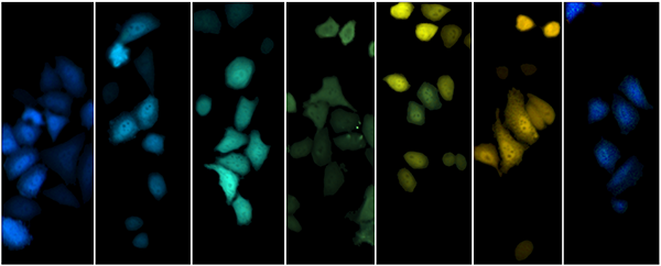
New Bioluminescent Phasor Imaging Technology Could Shine Light on Immune Functions

Aug. 2, 2022 - UC Irvine researchers Michelle Digman and Jennifer Prescher have developed a new technology for multiplexed imaging at the microscale using bioluminescent probes. The engineered microscopy platform will allow scientists to simultaneously track and manipulate multiple kinds of cells and molecules over prolonged periods, helping them better understand the immune system and how it dovetails with metabolism. The research was published in Nature Methods in June 2022.
“Being able to track and globally probe immune cells and their metabolic functions all at once, over time, in a living animal, will help with diagnosis and treatment of diseases such as cancer and neurodegeneration,” said Digman.
The two researchers, Digman, associate professor in biomedical engineering, and Prescher, professor in chemistry, came together for this interdisciplinary collaboration at the initiation of a graduate student, now alumnus, Zi Yao ’20 (Ph.D.) When Yao was earning his doctorate in chemistry at UCI under the advisement of Prescher, he took a graduate level course in molecular and cellular biophotonics, taught by Digman.
Yao asked Digman if they could try doing phasor analysis, a method used to separate colors, with bioluminescence data, the light that is produced by a living organism. Light-emitting enzymes have been engineered to produce various colors that help researchers observe more than one type of cell in tissues or more than one function in the cell. The phasor analysis is a graphical plot that provides the separation of colors without relying on computationally intensive processes to unmix colors. Yao is a co-author on the paper, along with chemistry alumnus Caroline Brennan ‘22 (Ph.D.) and biomedical engineering postdoctoral scholar Lorenzo Scipioni.
The two labs worked together to develop the bioluminescent phasor technology that would enable simultaneous detection of spectral emissions from multiple bioluminescent probes.
“We can differentiate up to six distinct bioluminescent reporters, but we are working on doing more and applying the phasor approach to imaging in live tissues,” said Prescher. “With new advances in low light photo detectors, or cameras that are extremely light sensitive, we will be able to pick up weaker signals and ‘see’ multiple processes relevant to immune function and other processes.”
The merger of bioluminescence with phasor analysis fills a long-standing void in imaging capabilities, and will bolster future efforts to visualize biological events in real time and over multiple length scales.
Prescher has pioneered methods to engineer bioluminescent probes and sensors, and Digman has many years of experience in optical imaging and applying phasor technology to various biological systems, including developmental biology, neurodegenerative diseases and cancer biology.
“It was really a perfect match between our two labs,” said Digman. “Our multidisciplinary expertise made for an excellent opportunity for us to work together.”
Digman and Prescher received funding for this research from The Paul G. Allen Frontiers Group through the Distinguished Investigator grant in 2020.
– Lori Brandt
