NIH Grant to Fund New ELCACT Instrument

Sept. 5, 2019 - This fall, a new state-of-the-art, highly sensitive microscope will be installed at UC Irvine’s Edwards Lifesciences Center for Advanced Cardiovascular Technology, giving researchers the capability to expand and accelerate their research. The instrument is funded in part by a highly competitive S10 equipment grant from the National Institutes of Health’s Office of Research Infrastructure Programs. UCI’s Office of the Vice Chancellor for Research provided matching funds.
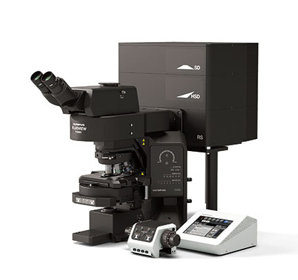
The $594,000 grant will fund an Olympus Fluoview FV3000 with MPM System, a multispectral, multiphoton laser scanning microscope. The tool will open the door to new cardiovascular and tissue engineering possibilities at ELCACT, a center that seeks to better understand cardiovascular disease and tissue vascularization. “This will allow us to conduct research and science related to topics such as heart cell structure-function-force generation relationships, macrophage mechanobiology, and the mechanics of growing vessels into implanted medical devices,” said principal investigator Elliot Botvinick, ELCACT associate director and professor in the departments of biomedical engineering and surgery. Botvinick also serves on the faculty at the Beckman Laser Institute.
Securing the grant was a five-year team effort led by Botvinick, Associate Professor Anya Grosberg and ELCACT Assistant Director Ann Fain. The effort included many biomedical engineering faculty who will use the equipment, including Tim Downing, Christopher Hughes, Abe Lee, Zhongping Chen, Arash Kheradvar, Kerry Athanasiou and Wendy Liu. “This showcases how center members were all instrumental in securing this grant,” Botvinick says.
The center’s current microscopes do not allow for three-dimensional views or optical sectioning of tissue. In contrast, the new microscope will have confocal and two-photon imaging, and on-stage incubation for longitudinal studies. Some of these studies utilize environmentally sensitive tissues that cannot be transported out of the building without compromising the research. The new instrument will be housed adjacent to the center’s Core Tissue Culture Facility, opening avenues for research involving mechanically sensitive samples.
“The microscope will greatly aid research in the areas of cardiac tissue engineering, vascularized on-a-chip platforms, cardiac valve tissue engineering, orthopedic tissue engineering” and more, Botvinick said. “It will provide critical state-of-the-art imaging capabilities not currently available at the center to accelerate and enhance our research.”
- Anna Lynn Spitzer
