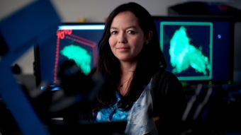
 A fluorescent green limb pokes outward from a cell wall under a high-powered microscope. The filament is loaded with VP40, an essential protein in the Ebola virus. The microscope is capturing it budding out in real time. It’s followed by another and another.
Those green protrusions may be the means by which the deadly virus races from cell to cell in humans, killing up to 60 percent of those infected, according to assistant professor of biomedical engineering Michelle Digman. She and fellow researchers at UC Irvine’s Laboratory for Fluorescence Dynamics and elsewhere have been using pioneering technology to meticulously track the molecular workings of the Ebola protein – and were the first to capture the action live.
Their hypothesis is that the filaments may act as needles that prick neighboring cells and inject the toxic material, a process that quickly goes viral. If that proves true, it could be key to the development of effective inhibitor treatments.
“This is the protein that seems to be really important for the final stage of the Ebola cycle, in which the virus leaves the cell that is infected in order to infect other cells,” says doctoral student Carmine Di Rienzo.
Digman and her co-researchers are among the few who were studying Ebola long before the current outbreak. In 2011, Ghanaian-born biochemist Emmanuel Adu-Gyamfi visited her UCI lab. He had seen BBC stories about Ebola as a child and – then a student at the University of Notre Dame – wanted to take advantage of new technology that lets scientists study the dynamics of living cells.
“We like challenges,” Digman says. “Emmanuel came to us and said, ‘I want to look at how this particular protein interacts in the cell. Can you do this?’ and we said, ‘Absolutely, yes.’”
As recently as a decade ago, it wasn’t possible to view live cells under microscopes. Scientists pored over fixed dead ones to puzzle out clues about their behavior. But a new technique called fluorescence dynamics allows researchers to track cells as they grow by tagging them with bright inks. Three people won the Nobel Prize in chemistry this year for their development of super-resolved fluorescence microscopy.
Still, it isn’t easy to trace the progress of the infinitesimally small viral particles. Digman and her colleagues created three-dimensional models and now compress hours of raw data to track VP40. There’s no risk from the isolated protein in the cell culture rooms; it needs the disease genome and six other proteins to fully form infectious Ebola.
The researchers’ work has gained critical importance with the re-emergence of Ebola in West Africa and the first confirmed deaths from the virus in the U.S. They recently published a paper in the journal Viruses that builds on their previous findings.
Usually, cells can absorb viruses and other harmful substances and neutralize them with acidic lipids. Not so with Ebola. Digman and the others have discovered that VP40 multiplies unimpeded inside the cell until finally the infected body’s membrane bulges out under the accumulated weight, budding into the needle-like filaments.
They’ve learned that the protein hijacks the cell’s motor to form the protrusions. They’ve seen detached filaments floating outside cells. Now they just need to see them pricking other cells. Or find that they do not and move on to other theories.
“Everything we learn about the Ebola VP40 protein is a surprise. It dies down, and then it comes back. It’s happened before, and it will happen again. Understanding these kinds of processes can help other scientists come up with targets to stop the spread,” Digman says
-Janet Wilson
