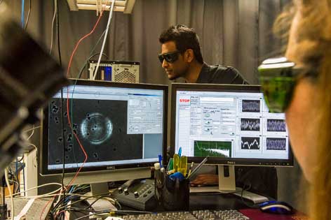
Biomedical engineer Elliot Botvinick uses optical tweezers to understand how disease takes hold
 At the intersection of engineering, physics, biology and medicine, Elliot Botvinick uses laser technology to study the molecular activity of diseases. Specifically, he utilizes optical tweezers, which let researchers hold and manipulate single molecules within cells. The work has myriad applications — in cancer, heart disease and diabetes, for example — and could lead to healthcare advances.
The old-fashioned “silo” structure of academic research does not apply to Botvinick, a UC Irvine assistant professor of biomedical engineering. He’s affiliated with The Edwards Lifesciences Center for Advanced Cardiovascular Technology on campus and is part of a multidisciplinary team of researchers at UCI’s Beckman Laser Institute & Medical Clinic.
A: In 2001, after receiving my doctorate in bioengineering at UC San Diego, I was awarded an Arnold and Mabel Beckman Foundation postdoctoral fellowship to work in the laboratory of BLI founder Michael Berns. At his request, I built a robotic microscope that was controllable through the Internet; it included optical tweezers. Through the development of this robot, I began to appreciate the potential of optical tweezers in the study of biology. At first, we worked with the San Diego Zoo’s Center for the Reproduction of Endangered Species to study the swimming force and energetics of primate sperm across species. I then used optical tweezers to explore the role of mechanical forces in cellular signal transduction, homeostasis and disease. This field, mechanobiology, ignited my love affair with the technology and represents the majority of the work in my laboratory at UCI.
Q: Simply put, how do optical tweezers work?
A: In “Star Trek,” we see spaceships trapped and manipulated by a focused energy beam. The conception and theory of optical tweezers are surprisingly similar. We can precisely measure forces acting on an optically trapped sphere, or “microbead,” that we either introduce into the cell or place in contact with molecules at the cell surface. My lab has integrated optical tweezers into a system also capable of 3-D imaging. Together, we can measure the unfolding of single molecules responsible for many cancers, study the effects of tissue stiffness on cancer metastasis and simulate vascular blood flow — all with real-time fluorescence imaging of a cell’s molecular responses.
Read more of the interview here>>
